
What Happens During Common Breast Imaging Procedures
Breast imaging plays a vital role in the early detection, diagnosis, and monitoring of breast conditions, including cancer. Understanding the different imaging procedures available helps patients and healthcare professionals make informed decisions about care and next steps. Here’s a breakdown of the most commonly performed breast imaging techniques and what they involve.
Why It’s Important to Understand Breast Imaging Procedures
For many, breast imaging can feel unfamiliar or daunting. Understanding what happens during these procedures, whether it’s a routine mammogram or a follow-up ultrasound, helps reduce anxiety, encourages informed decision-making, and improves the overall patient experience.
From a clinical standpoint, different imaging techniques are chosen based on individual risk factors, symptoms, or previous findings. A clear understanding when breast imaging is recommended and what it involves ensures that each step is aligned with the diagnostic goal, whether it’s early detection, clarification of abnormalities, or guiding further care.
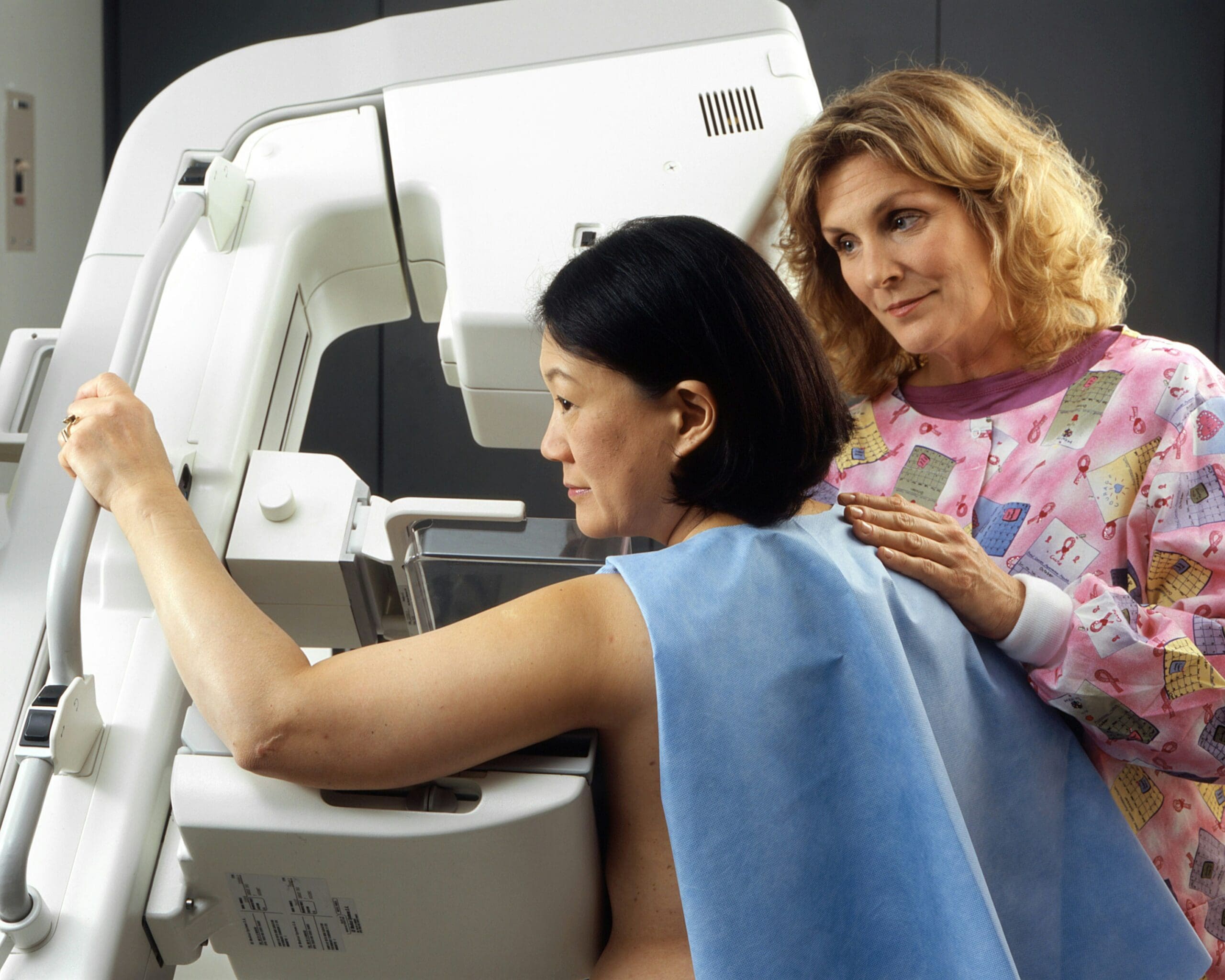
Mammography: The Primary Screening Tool
Mammography is one of the most established breast imaging techniques, using low-dose X-rays to capture detailed images of breast tissue. It is primarily used as a screening tool for individuals without symptoms, but also serves diagnostic purposes when signs such as lumps or nipple discharge are present. The process involves compressing each breast between two plates to spread the tissue evenly — a step that may cause brief discomfort but is essential for image quality. The examination typically takes around 20 minutes, and results are generally available within a few days.
As a first-line modality, mammography often guides subsequent imaging decisions. When findings are unclear or further assessment is needed, clinicians may recommend an ultrasound or an MRI to obtain additional information. Clinical factors, including age, risk level, and physical findings, influence this process.
Breast Ultrasound: Gives A Closer Look at Abnormalities
Breast ultrasound is frequently used alongside mammography, particularly for individuals with dense breast tissue, where X-ray images may not offer sufficient clarity. It employs high-frequency sound waves to generate real-time visuals of the internal structures of the breast, allowing clinicians to differentiate between solid masses and fluid-filled cysts more effectively.
This technique is non-invasive and free from radiation exposure, making it suitable for repeated use. During the scan, a conductive gel is applied to the skin, and a handheld transducer is moved across the breast to capture images from various angles.
Breast MRI: For High-Risk and Complex Cases
Magnetic Resonance Imaging (MRI) of the breast uses powerful magnets and radio waves to produce highly detailed, cross-sectional images. It’s typically reserved for high-risk individuals, such as those with known BRCA1 or BRCA2 mutations, or a strong family history of breast cancer, and for cases where other imaging results are inconclusive.
The procedure involves lying face down in an MRI scanner for up to an hour. A contrast agent is often injected intravenously to enhance image detail. While MRI is more sensitive than other techniques, it can also yield false positives, so it’s generally used as part of a carefully considered diagnostic plan.
Tomosynthesis: 3D Mammography for Better Clarity
Also known as 3D mammography, digital breast tomosynthesis (DBT) captures multiple images of the breast from different angles and reconstructs them into a three-dimensional format. This improves the visualisation of overlapping tissues and enhances the detection of small abnormalities.
The experience is nearly identical to a standard mammogram in terms of preparation and duration. Still, the resulting images provide greater depth and precision — particularly valuable for individuals with dense breasts or complex tissue structures.
Bringing Clarity to a Complex Process
Breast imaging procedures play a crucial role in both routine screening and targeted diagnosis. While the techniques may differ in method and purpose, they all contribute to building a clearer picture of breast health. By understanding what each procedure involves, patients and professionals alike are better equipped to make informed, confident decisions at every stage of care. A comprehensive women’s health checklist can further support this process, ensuring that breast imaging is integrated with other preventive screenings and wellness practices for a more complete approach to care.